Filed under: Uncategorized | Comments Off on My Backyard Today
Interesting Science Picture XVIII: Spiders Eat Bats
Call me disturbed.
 (Source)
Figure 2. Bats caught by spiders. A – Adult female Avicularia urticans feeding on a Greater Sac-winged Bat (Saccopteryx bilineata) on the side of a palm tree near the Rio Yarapa, Peru (photo by Rick West, Victoria, Canada; report # 1). B – Adult Proboscis Bat (Rhynchonycteris naso) entangled in a web of Argiope savignyi at the La Selva Biological Station, northern Costa Rica (photo by Mirjam Kno¨ rnschild, Ulm, Germany; report # 14). C – Dead bat (presumably Centronycteris centralis) entangled in an orb-web in Belize (photo by Carol Farneti-Foster, Belice City, Belize; report # 12). D – Dead bat (Myotis sp.) entangled in a web of Nephila clavipes in La Sirena, Corcovado National Park, Costa Rica (photo by Harald & Gisela Unger, Ko¨ ln, Germany; report # 17). E – A bat caught in the web of an araneid spider (possibly Eriophora sp.) in Tortuguero National Park, Costa Rica (photo by Cassidy Metcalf, USA; report # 18). F – Live bat trapped in web of Nephilengys cruentata in a thatch roof at Nisela Lodge, Swaziland (photo by Donald Schultz, Hollywood, USA; report # 47). G – Volant juvenile Proboscis Bat (Rhynchonycteris naso) entangled in web of Nephila clavipes photographed in a palm swamp forest near Madre de Dios, Peru (photo by Sam Barnard, Colorado Springs, USA; report # 7). H – Dead bat entangled in web of a female Nephila clavipes in tropical rainforest in the middle of the Rio Dulce River Canyon near Livingston, Guatemala (photo by Sam & Samantha Bloomquist, Indianapolis, USA; report # 11). I – Dead bat (Rhinolophus cornutus orii) caught in the web of a female Nephila pilipes on Amami-Oshima Island, Japan (photo by Yasunori Maezono, Kyoto University, Japan; report # 35). J, K – A small bat (superfamily Rhinolophoidea) entangled in web of Nephila pilipes at the top of the Cockatoo Hill near Cape Tribulation, Queensland, Australia (photo by Carmen Fabro, Cockatoo Hill, Australia; report # 39). The spider pressed its mouth against the dead, wrapped bat, indicating that it was feeding on it. A Nephila pilipes male also present in the web (K) may have been feeding on the bat as well. L – Dead vespertilionid bat entangled in the web of a female Nephila pilipes in the Aberdeen Country Park, Hong Kong (photo by Carol S.K. Liu from AFCD Hong Kong, China; report # 32).
Filed under: Bats, Spiders | Comments Off on Interesting Science Picture XVIII: Spiders Eat Bats
Interesting Science Picture XVII
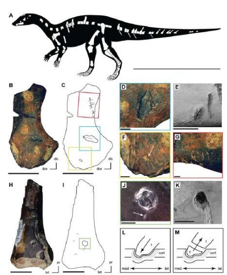
Evidence of a crocodyliform feeding on a juvenile ‘hypsilophodontid’ dinosaur:
 (Source) Figure 2. Feeding traces on juvenile ‘hypsilophodontid’ bones (Kaiparowits Formation) compared to those derived via actualistic experiments. A. Skeletal reconstruction of the undescribed ‘hypsilophodontid’ from the Kaiparowits Formation with known material shown in white (modified from [65]). B. Partial left scapula (UMNH VP 21104) with feeding traces collected from UMNH locality 303. C. Outline drawing of left scapula (UMNH VP 21104) with feeding traces highlighted and colored boxes showing the locations of figure parts D, F, and G (colors match the respective figure parts). D. Bisected pit on the left scapula (UMNH VP 21104). E. Bisected pit on a modern cow femur produced by Alligator mississippiensis during actualistic experiments [20]. F. Small pit (highlighted by white arrow) on the proximal portion of the left scapula (UMNH VP 21104). G. Series of small scores present along the ventral margin of the neck of the left scapula (UMNH VP 21104). H. Distal portion of a right femur (UMNH VP 21107) with feeding traces collected from UMNH locality 303. I. Outline of right femur (UMNH VP 21107) with feeding traces highlighted and colored box showing the location of figure part J. J. Puncture containing an embedded tooth present on the right femur (UMNH VP 21107) and a small pit (highlighted by white arrow) just ventral to the puncture. K. Puncture present on a modern cow femur produced by A. mississippiensis during actualistic experiments [20]. L. Reconstruction of the hypothesized impact of the crocodyliform tooth with the right femur, creating the puncture observed in UMNH VP 21107. M. Reconstruction of the hypothesized fracturing of the damaged crocodyliform tooth crown, resulting in the embedded tooth observed in UMNH VP 21107. Scale bar equals one meter in A, 10 mm in B, E, H, and K, 2 mm in D, F, G, and J. Abbreviations: cort, cortical bone; dis, distal; dor, dorsal; f, tooth fragment; lat, lateral; med, medial; pr, proximal; t, tooth crown. doi:10.1371/journal.pone.0057605.g002
I put hypsilophodontid in quotes for several reasons. First, there is good evidence to indicate the taxon is paraphyletic. Second, the authors of the paper the picture was taken indicate the specimens are from a previously undescribed taxon and refer to the specimen as the ‘Kaiparowits hypsilophodontid.’
Literature
Boyd et al (2013) Crocodyliform Feeding Traces on Juvenile Ornithischian Dinosaurs from the Upper Cretaceous (Campanian)
Kaiparowits Formation, Utah. PLoS ONE 8(2): e57605. doi:10.1371/journal.pone.0057605
Filed under: Science Pictures | Comments Off on Interesting Science Picture XVII


